ISLES Challenge 2018
Ischemic Stroke Lesion Segmentation

Looking for previous ISLES challenges? 2017, 2016, 2015


Important Notices

  1. The participants have been emailed to submit their proceedings abstracts until Oct. 15, 2018.
  2. Congratulations to this year's finalists! The list with the finalists and photos have been added.
  3. 10 August 2018: Due to an issue that has been fixed now in the evaluation scripts, we'd like to kindly ask you to re-upload your testing results. The deadline for submission has been extended to: August 17th. Let us know if you encounter any issues.
  4. 3 July 2018: The training set has been updated after fixing some cases having issues with Dicom-Nifti conversion.
  5. Training data is online on SMIR.
  6. ISLES will be held in conjunction with the BrainLes Workshop and the BraTS Challenge as a full-day satellite event of MICCAI 2018 on September 16, 2018.

Overview

Welcome to Ischemic Stroke Lesion Segmentation (ISLES) 2018, a medical image segmentation challenge at the International Conference on Medical Image Computing and Computer Assisted Intervention (MICCAI) 2018 (10-14th September). ISLES will be held jointly with the BrainLes Workshop and the BraTS Challenge.

This year ISLES 2018 asks for methods that allow the segmentation of stroke lesions based on acute CT perfusion data. Therefore, a new data set of 103 stroke patients and matching expert segmentations are provided. Please find more details about the challenge's motivation, data, rules and information about how to participate in the paragraphs below.

Results

The finalist have been awarded on the 16th September 2018 as follows:

Photos

Dates

14 June 2018 Registration opens and distribution of training data
23 July 2018 Deadline for submitting abstract
3 August 2018 Distribution of test data
10 August 2018 Early bird rate for MICCAI 2018 registration
17 August 2018 Deadline for submitting test data results
16 September 2018 Presentation of method and result on the workshop day
15 October 2018 Submission deadline for extended LNCS papers (8 to 12 pages)
23 October 2018 Reviews deadline for extended LNCS papers
5 November 2018 Camera-ready and signed copyright form submission deadline for extended LNCS papers

About

The ISLES Challenge

This challenge for stroke lesion segmentation has become very popular the past three years (2015, 2016, 2017) and yielded various methods that help to tackle important challenges of modern stroke imaging analysis. This year the challenge provides acute stroke CT perfusion imaging scans and manually outlined core lesions on MRI DWI scans acquired soon thereafter.

How the off-site challenge works

If you are interested in participating, you are invited to download the training set, including both CT perfusion scans as well as the corresponding expert segmentations of the infarct lesions. This will allow you to validate and optimise your method as much as you favour.

Shortly before MICCAI 2018 will take place, a set of test cases will be released of which participants will be asked to run their algorithm on and upload their segmentation results in form of binary image maps. To complete a successful participation, participants will need to submit an abstract, describing the employed method.

The organizers will then evaluate each case and establish a ranking of the participating teams. All results will be presented during a satellite event at MICCAI 2018 and will be discussed with invited experts and all workshop attendees.

Each team will have the opportunity to present their submitted method as a poster, while selected teams will be asked to give a brief presentation detailing their approach. Eventually, submissions will be included in the workshops post-proceedings and potentially compiled for a high-impact journal paper to summarise and present the findings.

Motivation

Clinical incentive

Defining location and extent of irreversibly damaged brain tissue is a critical part of the decision-making process in acute stroke, as demonstrated by the recent DAWN and DEFUSE-3 trials. MRI using diffusion and perfusion imaging can be used to distinguish between infarcted tissue ("core") and hypoperfused lesion tissue ("penumbra"). More recently, CT has been used to triage stroke patients, because of its speed, availability, and lack of contraindications. However, it is challenging to identify irreversibly damaged tissue (core) on CT studies. It has been proposed that CT perfusion could be used for this purpose, and many commercial softwares strive to perform this measurement. However, such automated methods are sparsely used and may be too simple to capture the full complexity of the data set. Therefore, there is a great need for advanced data analysis techniques that could help to define these regions for diagnosis in a more reproducible and accurate way and eventually support clinicians in their decision-making process (e.g., deciding for or against thrombolytic therapy). The MRI studies in this challenge were acquired immediately after the CT scans and therefore the region of DWI abnormality can act as a gold standard for irreversible brain infarction.

Why participate in this challenge?

Medical image processing comprises many tasks, for which new methods are regularly proposed. However, varying data set size and heterogeneity make it nearly impossible to compare different approaches in a fair way. By providing a high-quality data set publicly and pre-defined evaluation rules, challenges like ISLES aim to overcome these limitations and create a common framework for adequate comparison of results.

Data

PATIENT COHORT

Imaging data from acute stroke patients in two centers who presented within 8 hrs of stroke onset and underwent an MRI DWI within 3 hrs after CTP were included.

ACUTE IMAGING DATA DETAILS

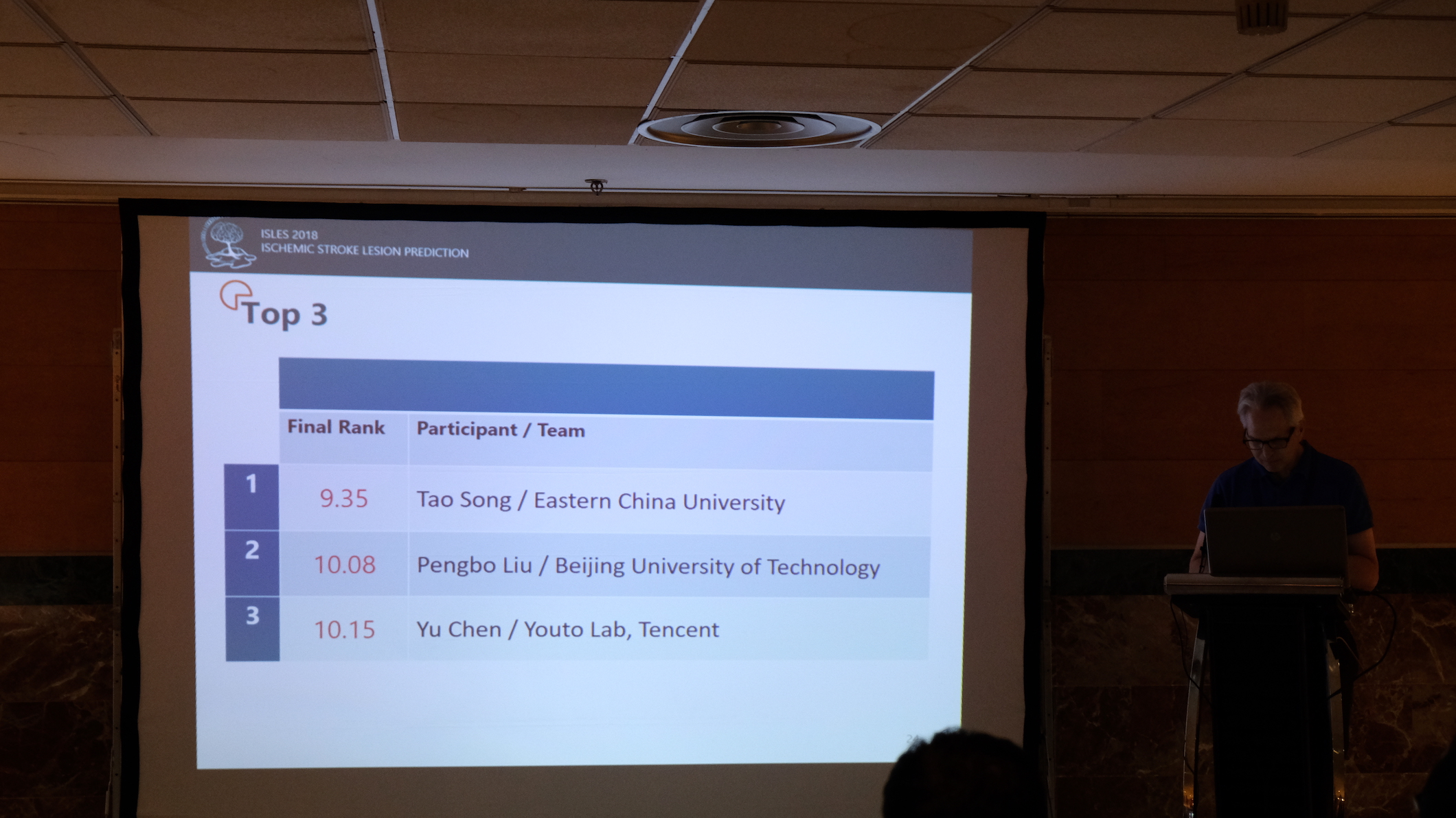
Training data set consists of 63 patients. Some patient cases have two slabs to cover the stroke lesion. These are non-, or partially-overlapping brain regions. Slabs per patient are indicated with letters "A" and "B" for first and second slab, respectively. The mapping between case number and training name is also provided at SMIR (e.g. Train_40_A = case 64; Train_40_B = case 65). Developed techniques will be evaluated by means of a testing set including 40 stroke cases. Acquired modalities are described in detail below.

GOLD STANDARD: DIFFUSION MAPS (DWI)

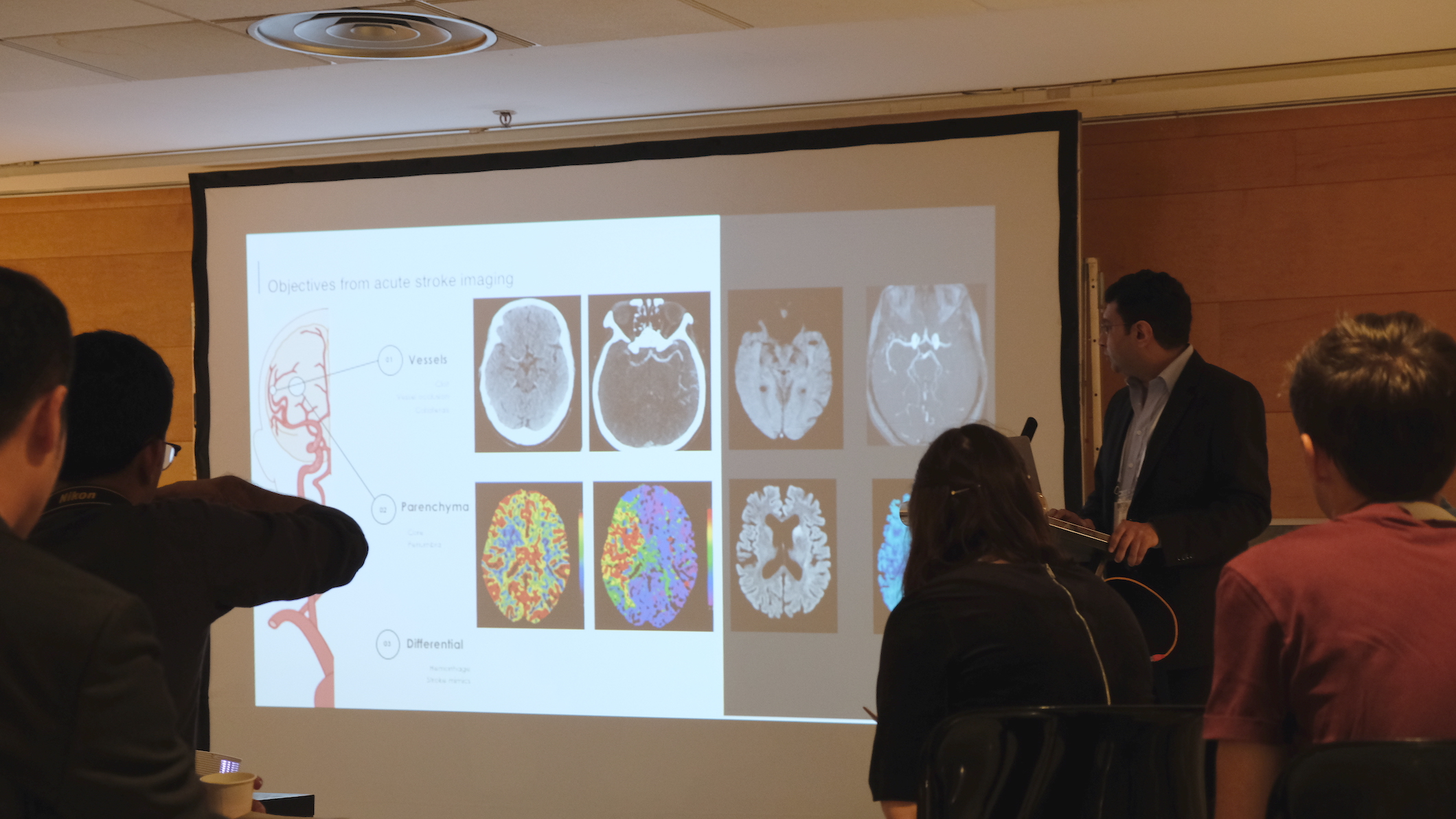
Infarcted brain tissue can be recognised as hyperintense regions of the DWI trace images (DWI maps). Provided ground-truth segmentation maps were manually drawn on those scans.

PERFUSION MAPS (CBF, MTT, CBV, Tmax, CTP source data)

To assess cerebral perfusion, a contrast agent (CA) is administered to the patient and its temporal change is captured in dynamic scans acquired 1-2 sec apart. Subsequently, perfusion maps are derived from these raw data for clinical interpretation. Different maps aim to yield different information, and the most commonly calculated maps include cerebral blood volume (CBV), cerebral blood flow (CBF), and time to peak of the residue function (Tmax). These perfusion maps serve as input to the algorithms.

PRIVACY AND DATA COPYRIGHT

By registering, each team agrees to use the provided data only in the scope of the workshop and neither pass it on to a third party nor use it for other publications. After the workshop takes place, the data will be released under a research license. No copyright transfer of any kind will take place, except in the case of a contribution to the LNCS post-proceedings special issue.

DOWNLOAD DATA

For data access and result submission, please register at SICAS Medical Image Repository:

Register

Please cite the following articles if you use ISLES18 data:

Cereda, Carlo W., Søren Christensen, Bruce CV Campbell, Nishant K. Mishra, Michael Mlynash, Christopher Levi, Matus Straka et al. "A benchmarking tool to evaluate computer tomography perfusion infarct core predictions against a DWI standard." Journal of Cerebral Blood Flow & Metabolism 36, no. 10 (2016): 1780-1789.

Hakim, Arsany, Søren Christensen, Stefan Winzeck, Maarten G. Lansberg, Mark W. Parsons, Christian Lucas, David Robben, Roland Wiest, Mauricio Reyes, and Greg Zaharchuk. "Predicting Infarct Core From Computed Tomography Perfusion in Acute Ischemia With Machine Learning: Lessons From the ISLES Challenge." Stroke (2021): STROKEAHA-120.

Rules

What is required to participate?

Each team wishing to participate in the ISLES challenge is required to:

  1. Register at the data distribution and evaluation platform (see participate for more details).
  2. Submit an abstract describing their method and their results on the training data set (see abstract).
  3. Upload the test data results before the deadline (see dates).
  4. Register to the associated MICCAI 2018 workshop and pay the attendance fee.
  5. Be present at the workshop with at least one team member.

Which methods are called for?

Any automatic method that predicts the lesion outcome is of interest. There is no restriction on new, innovative, or unpublished methods. Semi-automatic methods are eligible for participation and will appear on ranking board, but will not be part of the competition, as it is impossible to rate the influence of the manual steps in a fair manner.

Abstract

ISLES requires the participating teams to submit an abstract of one page in LNCS format, which will be reviewed by the organizers. The text will be distributed among the MICCAI 2018 attendees on the MICCAI 2018 USB drive and uploaded to the challenge result web-page. Accepted manuscripts will be also published in Springer Lecture Notes in Computer Science (LNCS) together with the proceedings of the Brainles workshop. See dates for the associated deadlines.

Requirements


Review


Submission


Extended Abstract for BrainLes Proceedings

Participate

Data distribution, registration and automatic evaluation will be handled by the SICAS Medical Image Repository:

Participate in ISLES


There you will find explanations on how to register, how to download the data, and how as well as in which format to upload your results. Furthermore, the evaluation scores obtained by each team will be listed there.

Organizers

This challenge is organized by:

Dr. Arsany Hakim, Inselspital Bern, Switzerland.

Dr. Mauricio Reyes, University of Bern, Switzerland

Prof. Roland Wiest, Inselspital Bern, Switzerland

Dr. Maarten G Lansberg, Stanford University, USA

Dr. Søren Christensen, Stanford University, USA

Dr. Greg Zaharchuk, Stanford University, USA

Stefan Winzeck, University of Cambridge, UK

David Robben, University of Leuven, Belgium

Contributors

Christian Lucas, University of Lübeck, Germany