ISLES Challenge 2016
Ischemic Stroke Lesion Segmentation

Looking for ISLES 2015? Click here.


Important Notices

  1. The final rankings can be found under results. We would like to thank all the participants for their hard work as well as the invited speakers for their interesting talks. The challenge has been a great success!
  2. We now provide a short FAQ sheet with the most common questions received.
  3. Information: Proceedings, on-site schedule
  4. Links: evaluation code, MICCAI registration
  5. See VSD for registration, training data download, test data download and evaluation system.
  6. ISLES 2016 joins with BRATS (tumor) and mTOP (traumatic brain injury) under the roof of the BrainLes workshop as a whole day event.

Overview

Welcome to Ischemic Stroke Lesion Segmentation (ISLES) 2016, a medical image segmentation challenge at the International Conference on Medical Image Computing and Computer Assisted Intervention (MICCAI) 2016 (October 17-21st).

ISLES 2016 provides a segmentation and a regression task:

Task I: Lesion outcome prediction

Task II: Clinical outcome prediction

A public dataset of diverse ischemic stroke cases and a suitable automatic evaluation procedure will be made available. The provided data allows the participating methods to combine image and clinical information in any combination to achieve the tasks' goals.

On this page, you can find details on how the challenge works in the about section, the clinical and research motivation behind it, the rules of how to participate, the data description and the evaluation scheme.

Results

The frozen ranking of the teams who participated at the ISLES workshop, held the 17th of October 2015 at the MICCAI 2016, is presented here and constitutes the final outcome of the challenges. Running results of the continued challenge can be found on the VSD data/evaluation web-page. Please note that the results there might vary from the ones presented in this table, as at the VSD page, the ASSD and HD are computed over all but empty segmentation masks, irregardless of the DC score.

TASK I

rank2 first author (team name) & affiliation cases ASSD1 DC HD1
3.34 Han-Byul Lee (KR-SUL)3
Department of Statistics, Seoul National University, Korea/span>
18/19 6.38±4.18 0.31±0.24 34.09±21.08
3.42 Yongchan Kwon (KR-SUK)3
Department of Statistics, Seoul National University, Korea
18/19 6.23±3.55 0.31±0.24 32.84±15.68
3.68 Youngwon Choi (KR-SUC)3
Department of Statistics, Seoul National University, Korea
18/19 6.46±4.38 0.30±0.25 37.70±21.61
4.38 Richard McKinley (CH-UBE)
University of Bern, Switzerland
19/19 7.10±4.19 0.26±0.19 44.30±24.64
4.60 Oskar Maier (DE-UZL)
Institute of Medical Informatics, Universität zu Lübeck, Germany
18/19 7.57±4.17 0.30±0.23 36.88±16.84
4.98 Haocheng Shen (UK-CVI)
CVIP, Comp. at School of Science and Eng., University of Dundee, UK
18/19 7.56±4.34 0.29±0.23 35.91±17.04
6.03 Lequan Yu (HK-CUH)
Dept. of Comp. Science and Eng., The Chinese University of Hong Kong
19/19 10.56±5.92 0.27±0.22 59.44±38.27
6.22 Qaiser Mahmood (PK-PNS)
Pakistan Institute of Nuclear Science and Technology, Islamabad, Pakistan
19/19 9.12±5.46 0.26±0.22 78.04±18.11
7.97 Ramandeep Randhawa (US-SFT)
University of Southern California, Fractal Analytics, TopicIQ
18/19 13.96±6.14 0.19±0.20 76.28±22.61

TASK II

rank4 first author (VSD-name) & affiliation cases avg abs error
1.5 Oskar Maier (DE-UZL)
Institute of Medical Informatics, Universität zu Lübeck, Germany
19/19 1.05±0.62
1.6 Han-Byul Lee (KR-SUL)3
Department of Statistics, Seoul National University, Korea/span>
19/19 1.10±0.70
1.8 Yongchan Kwon (KR-SUK)3
Department of Statistics, Seoul National University, Korea
19/19 1.26±0.81
2.3 Youngwon Choi (KR-SUC)3
Department of Statistics, Seoul National University, Korea
19/19 1.37±1.00
2.5 Qaiser Mahmood (PK-PNS)
Pakistan Institute of Nuclear Science and Technology, Islamabad, Pakistan
19/19 1.26±0.87

1Computed excluding the failed cases (where DC=0, see cases-column), therefore not suitable for a direct comparison of methods
2Relative rank, computed as described below in the evaluation section
3These three submissions are variants of a single method and count as one for award purposes.
4Average if case-wise ranks. Does not necessary reflect the average absolute error.

Each row is linked to a method description as PDF file. The complete proceedings can be found here (v2016-09-27)

Photos


Fig.: Click on any to start slide-show. Photos by Oskar Maier.

Conclusion

The BrainLes workshop took place at the 17th of October 2016. In front of a full room of participants and interested researchers from all over the world, the first three workshop contributions were presented, followed by an invited talk from Christian Barillot. During the next session, the mTOP challenge was introduced, presented and the rankings revealed.

After the coffee break, it was time for the ISLES challenge. First, Roland Wiest promoted the neuroradiological perspective on stroke lesion segmentation and detailed the requirements arising from the field. Then we gave a short introduction of the challenges' tasks and announced the winners (Congratulations!). These then had the opportunity to present their methods in form of a short presentation. Finally, a first interpretation of the ISLES results was given.
In the afternoon, the workshop continued with invited lectures from George Stranjalis and Koen Van Leemput, followed by the BRATS challenge. After a few more participating works, a lively discussion arose, sprinkled with much welcome recommendations for a third edition of the challenge.

We would like to thank the participants for their interest and hard work, the invited speakers for their valuable contributions and the audience for their numerous appearance and the lively discussions. It has been a great experience with all of you!

About

Why challenges?

Medical image processing comprises a large number of tasks, for which new methods are regularly proposed. But, more often than desirable, it is difficult to compare their fitness, as the reported results are obtained on private datasets. Challenges aim to overcome these shortcomings by providing (1) a public dataset that reflects the diversity of the problem and (2) a platform for a fair and direct comparison of methods with suitable evaluation measures. Thus, the scientific progress is promoted (see grand-challenge.org for a detailed rationale behind challenges).

Past challenges

During the recent years, the need for challenges has been emphasized in the medical image processing community. Especially in the scope of the MICCAI and International Symposium on Biomedical Imaging (ISBI) conferences, a number of challenges have been already successfully organized. All of these are collected centrally at grand-challenges.org. Last year, we organized with ISLES 2015 a successful ischemic stoke lesion segmentation challenges with a similar task.

The ISLES 2016 challenge

The ISLES 2016 challenge treats the challenging problem of stroke lesion/clinical outcome prediction from acute MRI scans. To this end, we will provide acute MRI scans of a large number of acute stroke cases and associated clinical parameters. The associated ground truth is the final lesion volume (Task I) as manually segmented in 3 to 9 month follow-up scans, and the clinical mRM score (Task II) denoting the degree of disability.

How it works

Interested participants can download a set of training cases (data description) with associated expert segmentations of the stroke lesions to train and evaluate their approach. Shortly before MICCAI 2016 takes place, a set of test cases will be made available and the participants upload their segmentation results in the form of binary images (Task I) respectively their mRM score predictions (Task II) as well as a abstract describing their method. The organizers then evaluate each case and establish a ranking of the participating teams. At the MICCAI 2016 conference, the results will be presented in the scope of a workshop and the results discussed together with invited experts. Each team will have the opportunity to present their method as a poster and, furthermore, a number of selected teams will be asked to hold a small presentation detailing their method. A number of high ranking and particularly interesting approaches will be invited to contribute to the workshops LNCS post-proceedings volume. After the challenge, the organizers and participants will together compose a high-impact journal paper to publish their findings.

Motivation

The typical stroke treatment procedure involves the acquisition of brain images at some time-since-stroke (TSS). Depending on the these and other factors a treatment decision is made and an intervention takes place after a time-to-treatment (TTT). The success of this intervention is assessed via the standardized TICI score. After the effects stabilized, the final clinical outcome is determined with the mRM and the final lesion outcome by brain imaging.

The watchword of most stroke organizations is time is brain. The treating physician has to decide in each case individually, whether the risky intervention (be it thrombectomy or thormbolysis) is justified by the potential gain, based solely on the lesion appearance, the time-since-stroke (TSS) and her or his personal experience. A method to reliably predict the actual lesion and clinical outcome from the acute scans alone would provide invaluable evidence for the clinical decision finding process. By taking the time-to-treatment into account, the prediction can be readily adapted depending on the availability of the treatment facilities.

Rules

What is required to participate?

Each team wishing to participate in the ISLES challenge is required to:

  1. Register at the data distribution and evaluation platform (see participate for more details).
  2. Submit an abstract describing their method and their results on the training data set (see abstract).
  3. Upload the test data results before the deadline (see dates).
  4. Register to the associated MICCAI 2016 workshop and pay the attendance fee (possible right up until the day of the challenge).
  5. Be present at the workshop with at least one team member.

Which methods are called for?

All types of automatic methods producing the desired end-points are welcome. We specifically do not restrict the participation to new, innovative and unpublished methods, but invite research groups from all over the world to enter their existing method in the competition. Semi-automatic methods are eligible for participation and will appear in the ranking, but out of competition, as it is impossible to rate the influence of the manual steps in a fair manner.

Organizational structure

ISLES is organized as part of a larger, four-part, full-day workshop at the MICCAI 2016 with the following organizational structure:

The four events should be regarded as a single workshop and a single registration allows to visit all of these and to participate in them. Please note that each part has its own rules of active participation, which can be found on their respective web-pages. The workshop schedule can be found here. Note that the final ordering is still subject to change.

Additional participation in another challenge than ISLES

All challenges and the workshop treat brain injury. Double contributions are highly welcome and only one registration at the MICCAI 2016 workshop is required.

Usage of ISLES data for the Brain Lesion Segmentation (BrainLeS) workshop

Participants are free to use the training data from ISLES to evaluate methods submitted to BrainLeS, but are encouraged to also participate in the challenge too. In such case, they are not required to submit the abstract to ISLES, instead their (accepted) BrainLeS manuscript will suffice, as long as it states the results obtained on the training set.

Abstract

ISLES requires the participating teams to submit an abstract of one page in LNCS format, which will be reviewed by the organizers. The text will be distributed among the MICCAI 2016 attendees on the MICCAI 2016 USB drive and uploaded to the challenge result web-page. See dates for the associated deadlines.

Requirements


Review


Submission

On-site method presentation

Poster


Presentation

Post-proceedings publication (LNCS)

Some selected methods from the three challenges and the workshop will be called upon to contribute an article to the BrainLes workshops Springer LNCS post-proceedings special issue with a longer version of their article. The respective participants are informed during the challenge and given a few weeks after the MICCAI to compose their article, which will have to undergo another review cycle.

Submitting results for multiple methods

A team can submit results from multiple methods, but an extra registration to the MICCAI 2016 challenge is required for each additionally submitted result.

Privacy and data copyright

By registering, each team agrees to use the provided data only in the scope of the workshop and neither pass it on to a third party nor use it for other publications. After the workshop took place, the data will be released under a research license. No copyright transfer of any kind will take place, except in the case of a contribution to the LNCS post-proceedings special issue.

Data

Image format

Uncompressed Neuroimaging Informatics Technology Initiative (NIfTI) format: *.nii.

(I/O libraries: MedPy (Python), NiftiLib (C,Java,Matlab,Python), Nibabel (Python), Tools for NIfTI and ANALYZE image (Matlab), ITK (C++))

Pre-processing

All MRI sequences are skull-stripped and anonymization was performed. No other pre-processing, such as re-sampling or co-registration is performed. This allows each participant to employ their favorite algorithms. For the skull stripping of the structural images the stripTs tool was utilized. For the PWI images, a brain mask was created with the BET algorithm from FSL and the resulting mask was applied to the corresponding PWI image with the fslmath tool from FSL. Varying hand crafted parameters were chosen for the BET algorithm, depending on the respective MRI sequence. The decision of using two different algorithms for the skull stripping rests upon qualitative assessment.

To facilitate the participation, we decided to provide the data in two variants: A version with all sequences co-registered and skull-stripped with a common brain mask; and a version without co-registration and with individual skull-stripping performed. This enabled the participants to apply their favorite registration method, if desired.

Data details

For ISLES 2016, 35 training and roughly 40 testing cases will be made public and can be downloaded from the SMIR platform after registration.

Clinical parameter details

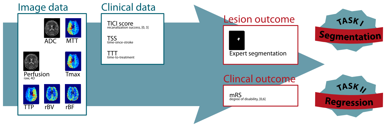
Thrombolysis in Cerebral Infarction (TICI) scale

The TICI scale, originally proposed by Higashida et al. (2003), provides a standardized method to evaluate intracranial perfusion assessed in cerebral angiography. Here, it is used to assess the achieve re-perfusion after a flow-restoration intervention such as thrombectomy.

Grade 0 No Perfusion. No antegrade flow beyond the point of occlusion.
Grade 1 Penetration With Minimal Perfusion. The contrast material passes beyond the area of obstruction but fails to opacify the entire cerebral bed distal to the obstruction for the duration of the angiographic run.
Grade 2 Partial Perfusion. The contrast material passes beyond the obstruction and opacifies the arterial bed distal to the obstruction. However, the rate of entry of contrast into the vessel distal to the obstruction and/or its rate of clearance from the distal bed are perceptibly slower than its entry into and/or clearance from comparable areas not perfused by the previously occluded vessel, eg, the opposite cerebral artery or the arterial bed proximal to the obstruction.
Grade 2a Only partial filling (2/3) of the entire vascular territory is visualized.
Grade 2b Complete filling of all of the expected vascular territory is visualized, but the filling is slower than normal.
Grade 3 Complete Perfusion. Antegrade flow into the bed distal to the obstruction occurs as promptly as into the obstruction and clearance of contrast material from the involved bed is as rapid as from an uninvolved other bed of the same vessel or the opposite cerebral artery.

Modified Rankin Scale (mRS)

The 90 days mRS is a standardized scale to assess the degree of disability 90 days after a stroke incidence and can be considered as the clinical outcome (van Swieten et al. (1988), see here for an online version of the test). The mRS score serves as endpoint for Task II.

Grade 0 No Symptoms at all.
Grade 1 No significant disability despite symptoms; able to carry out all usual duties and activities.
Grade 2 Slight disability; unable to carry out all previous activities, but able to look after own affairs without assistance.
Grade 3 Moderate disability; requiring some help, but able to walk without assistance.
Grade 4 Moderately severe disability; unable to walk without assistance and unable to attend to own bodily needs without assistance.
Grade 5 Severe disability; bedridden, incontinent and requiring constant nursing care and attention.
Grade 6 Dead.

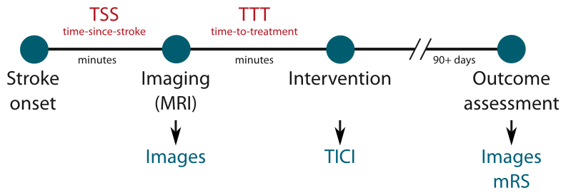
Time-since-stroke (TSS)

TSS is an important factor as only a fast re-establishing of the cerebral perfusion can lead to benefits for the patient. The less time passed since stroke onset, the more likely a re-perfusion can salvage brain tissue.

Time-to-treatment (TTT)

TTT denotes the time passed between imaging the patient and the re-perfusion treatment. Similar to the TSS, its value determines the amount of salvageable brain tissue.

Acute imaging details

For this challenge, MRI sequences recorded at the time of admission are as detailed below. For more information on the sequences, see e.g. http://mriquestions.com.

ADC map

Constructed from the DWI images, the ADC map show the underperfused area as hypointense region. Compared to the DWI trace images, the ADC map does not suffer from confounding T2 shine-through effects.

Raw perfusion data

Perfusion imaging (here: dynamic susceptibility imaging) denotes the perfusion of brain tissue with blood over time. The raw perfusion data are multiple 3D scans performed at equal time-intervals combined into a 4D volume.

Perfusion maps (MTT, Tmax, TTP, rBV, rBF)

For clinical assessment various maps can be computed from the raw perfusion data, each of which denotes different aspects of the measured brain perfusion. For ISLES 2016, a number of maps computed via block decomposition using the Olea Sphere software are provided.

Follow-up stroke imaging

To assess the final lesion outcome, an anatomical sequence is acquired when the stroke lesion stabilized. The manually segmented lesion in this scan constitutes the final lesion outcome and serves as end-point for Task I.

Participate

Data distribution, registration and automatic evaluation will be handled by the SICAS Medical Image Repository:

Participate in ISLES

There you will find explanations on how to register, how to download the data and how as well as in which format to upload your results. Furthermore, the evaluation scores obtained by each team will be listed there.

Evaluation

Task I: Lesion outcome

The evaluation comprises three evaluation measures, each of them highlighting different aspects of the segmentation quality. From the computed evaluation scores, the global ranking of each team is established as described on ISLES 2015 for the SISS task. See there under Evaluation for more details.

Task II: Clinical outcome

Details on the evaluation of the regression task will be provided soon.

Organizers

This challenge is organized by

Egger, Karl, Clinic for Neuroradiology & University Medical Center Freiburg, Germany.

Maier, Oskar, Institute of Medical Informatics, Universität zu Lübeck, Germany (directed by Prof. Dr. rer. nat. habil. Heinz Handels)

Reyes, Mauricio, Institute for Surgical Technology & Biomechanics, Universität Bern, Switzerland

Wiest, Roland, Department of Diagnostic and Interventional Radiology, Inselspital Bern, Switzerland.